
ProWay optical microscope and accessories
The ProWay biological optical is suitable for observation and digital image and video capture in the fields of cytology and microbiology.
Price
VAT included
ProWay optical microscope and accessories
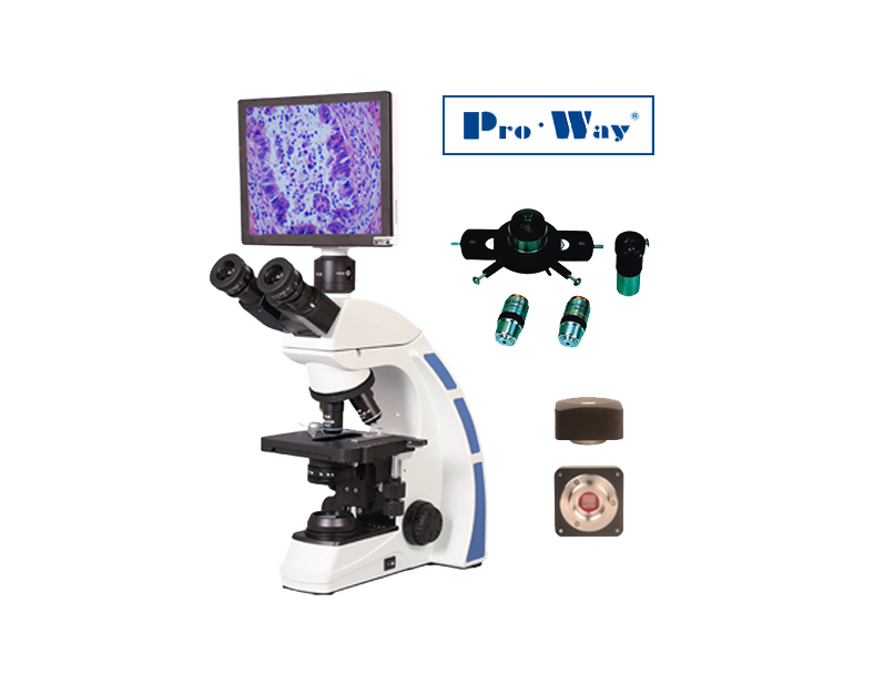
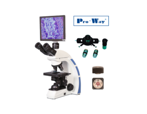
The ProWay biological optical ProWay is suitable for observation and digital image and video capture in the fields of cytology and microbiology. It comes with accessories for every need, such as:
- DCE-PW300 CMOS camera, with 3 MP resolution and a USB interface, for capturing images and video using ImageView / ToupView (Windows) or ToupLite (macOS / OS X, Linux),
- DCE-PW500 CMOS camera, with 5 MP resolution and a USB interface, for capturing images and video using ImageView / ToupView (Windows) or ToupLite (macOS / OS X, Linux),
- XCAM1080PHA camera, with 2.1 MP resolution, 1920x1080p video at 60 fps (HDMI), 1920×1080 at 30 fps (USB), HDMI and USB output interfaces, SD card slot, mouse, 4GB SD card, lens adapter, and power cable.
- TFT LCD Tablet with Built-in Camera 5 MP resolution (I/O interfaces: USB, MINI USB, SD CARD, MICRO HDMI),
- Phase Contrast Kit (20x PH, 100x PH),
- 20x, 40x (s), 60x, 100x, 100x (s, oil) objectives and corresponding PH objectives for phase contrast
The legislation on the advertising of medical devices (Legislative Decree 46/97, as amended by Legislative Decree 37/10) stipulates that access to medical information relating to medical devices is restricted to authorized persons, doctors, or other healthcare professionals. For further information on our products, please consult your doctor or pharmacist.
• ProWay XSZ-PW208 / XSZ-PW208T / XSZ-PW208LCD Biological Optical Microscope

ProWay XSZ-PW208 (binocular) and XSZ-PW208T (trinocular) Biological Optical Microscopes
| XSZ-PW208 | |
| Optical System | UIS – Infinite Optical System |
| Test | Trinocular, tilted 30°, interpupillary distance: 57–75 mm |
| Eyepieces | PL10X/20mm, retro-projection, wide-angle with diopter adjustment of +/- 5 degrees |
| Bridge | Reverse Quadruple |
| Objectives | Achromatic objectives with infinity focus [4x, 10x, 40x (s), 100x (s, oil)] |
| Coffee Table | Rectangular coaxial mechanical bridge, measuring 140 mm × 132 mm, with a travel range of 76 mm × 50 mm. |
| Focusing System | Macrometric and micrometric knobs, with limit stops and adjustable tension |
| Capacitor | Koehler A.N. 1.25 (with port for Phase Contrast and Dark Field kits ) |
| Lighting | Wide voltage range: 100V–240V; 3W LED lamp with dimmer (fixed center). |
| Optional Accessories | Trinocular head (XSZ-PW208T), LCD tablet (XSZ-PW208LCD), dark-field kit, phase-contrast kit (10x, 20x, 40x, 100x), DCE-PW300 CMOS digital camera, simple polarization (with analyzer and polarizer). |
• ProWay XSZ-PW208LCD Digital Optical Microscope

ProWay XSZ-PW208LCD Digital Optical Microscope
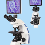
The enhanced XSZ-PW208LCD model integrates the XSZ-PW208T trinocular model with an LCD touchscreen tablet equippedwith a camera for capturing images and videos, featuring the following specifications:
| LCD Tablet | |
| Screen | 9.7-inch TFT LCD Touch Screen |
| Operating System | Android 4.2.2 |
| Interface | USB, Mini USB, SD memory card, Micro HDMI; can be connected to mice and keyboards via USB, and to monitors or projectors via Micro HDMI |
| Bedroom | 5.0 MP CMOS sensor chip |
| Software | TouchScope 2.5.6 |
| Connectivity | Wi-Fi – Bluetooth |
| Power Supply | 100-240V AC Power Supply |
| Accessories | Includes a C-mount adapter with 0.5X manual focus |
• Accessories
Phase Contrast Kit

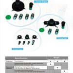
Phase Contrast Kit – Sliding Type
The Phase Contrast Kit consists of a 100x PH-I (or 20x PH-I) objective lens and a sliding module for the Koehler condenser (Sliding Type).
Objectives
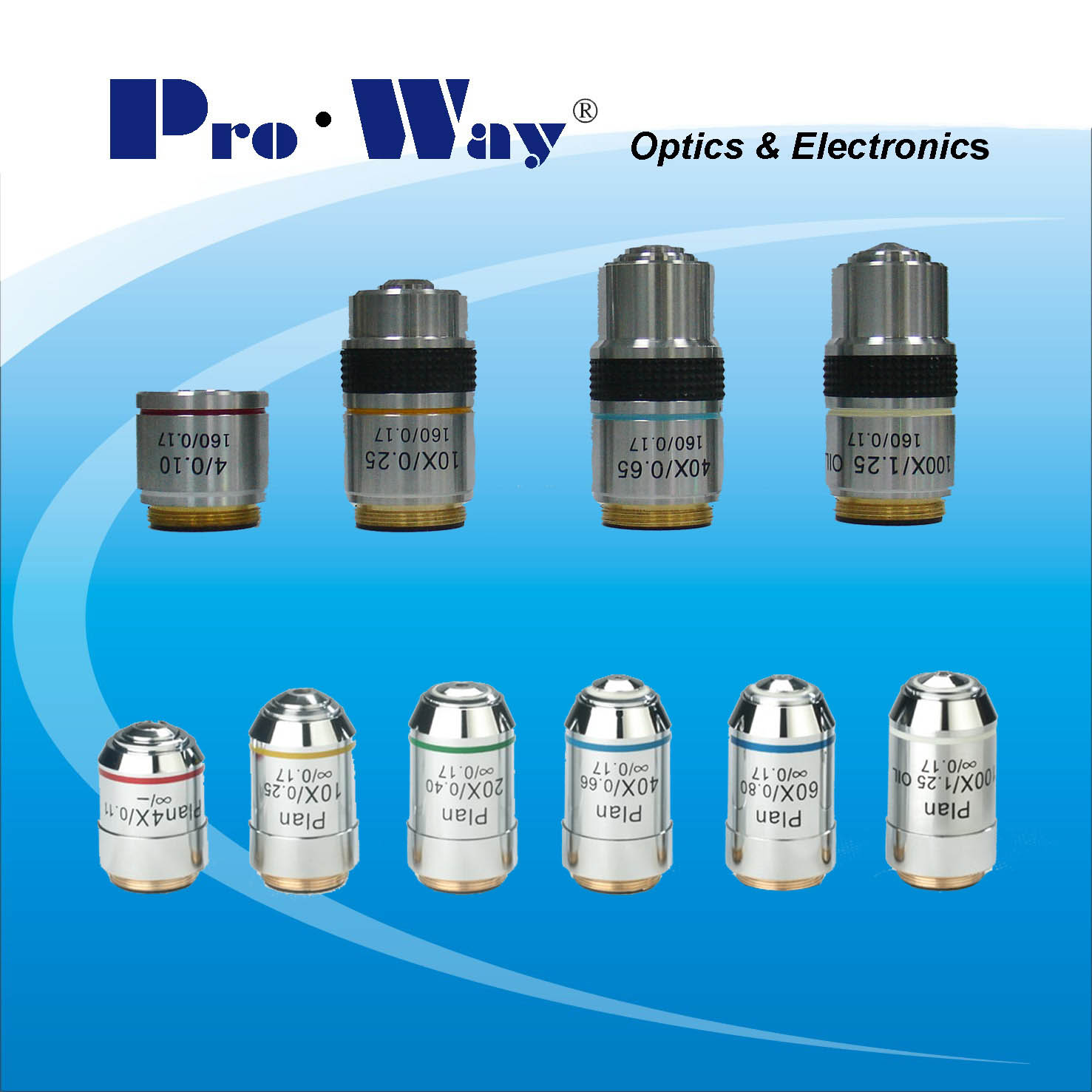
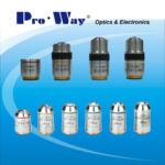
ProWay microscope objectives
Achromatic objectives: 4x, 10x, 20x, 40x, 60x, 100x (s, oil) and corresponding PH objectives for phase contrast.
DCE-PW300/DCE-PW500 CMOS Digital Camera
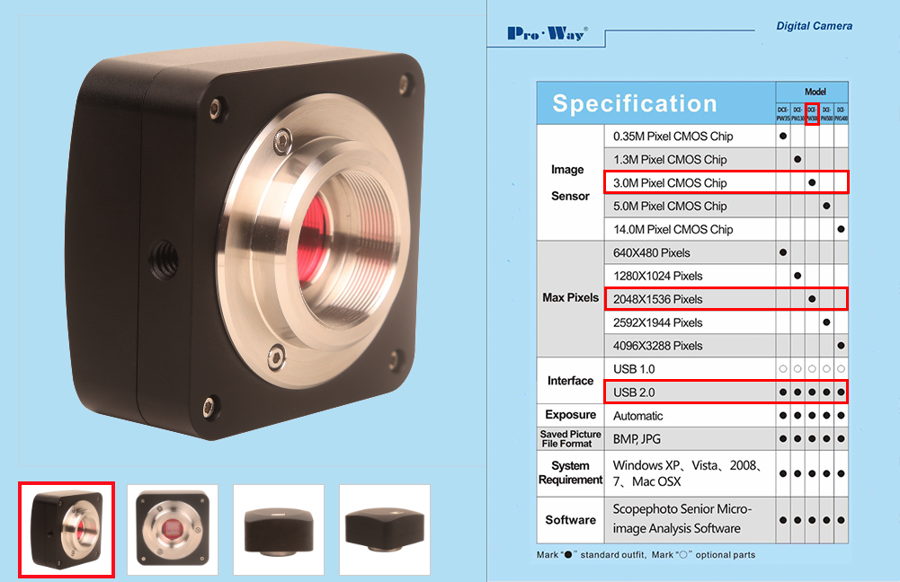
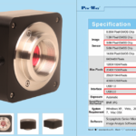
Proway DCE-PW300 CMOS Digital Camera

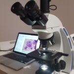
XSZ-PW208T with DCE-PW300/DCE-PW500 camera . On the left, ImageView software displaying a nasal cytology image.
DCE-PW300 Digital Camera
| DCE-PW300 | |
| Bedroom | 3.0 MP 1/2-inch CMOS sensor chip |
| FPS/Max. Resolution | 18@2048×1536 22@1024×768 43@680×510 |
| Interface | USB 2.0 |
| Exhibition | Automatic |
| Fire | Manual |
| File Format | BMP, JPG |
| Software | ImageView® (included with Windows) andToupLite(downloadable for Windows, macOS, and Linux) |
DCE-PW500 Digital Camera
| DCE-PW500 | |
| Bedroom | 5.0 MP 1/2.5″ (5.70 × 4.28) CMOS chip sensor |
| FPS/Max. Resolution | 5@2592×1944 18@1280×960 60@640×480 |
| Interface | USB 2.0 |
| Exhibition | Automatic |
| Fire | Manual |
| File Format | BMP, JPG |
| Software | ImageView® (included with Windows) andToupLite(downloadable for Windows, macOS, and Linux) |
XCAM1080PHA Digital Camera
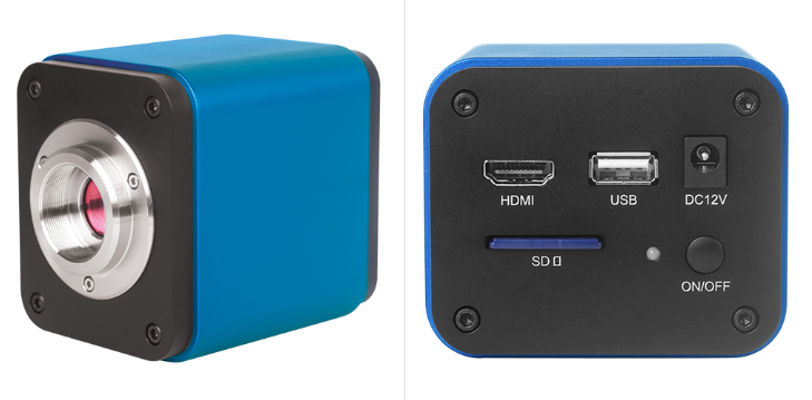
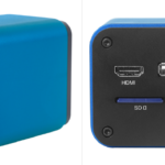
XCAM1080PHA HDMI Digital Camera
| XCAM1080PHA | |
| Bedroom | 2.1 MP 1/2.8″ (5.38 × 3.02) CMOS sensor chip |
| FPS/Max. Resolution | 60@1920x1080P (HDMI) 30@1920×1080 (USB) |
| Interface | USB 2.0, HDMI (out), SD card, Mouse |
| Exhibition | Automatic |
| Fire | Manual |
| File Format | BMP, JPG |
| Software | XCamView UI (built-in, via HDMI), ImageView (CD) /ToupView / ToupLite (downloadable) (Windows, macOS, Linux), Microsoft® Windows® XP / Vista / 7 / 8 / 8.1 / 10 (32- and 64-bit), OS X (Mac OS X), Linux (via USB) |
LCD tablet with a built-in 5MP camera and TouchScope / S-EYE software for Android
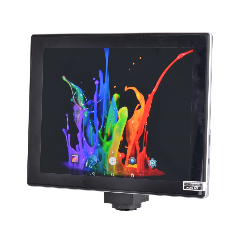
S-EYE 5MP LCD Tablet Camera with C-mount
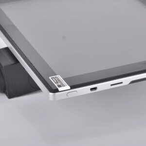
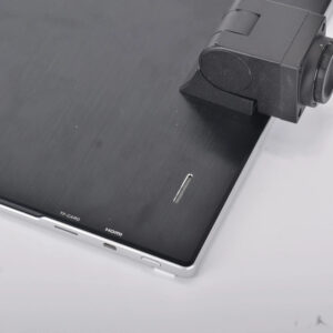
LCD Tablet I/O: mini-HDMI, TF Card, Power On/Off Button
|
LCD Tablet |
Screen | 1024×768 IPS LCD, 9.7” TFT Touch Screen |
| Operating System | Android 4.2.2 | |
| I/O Interfaces | USB/Mini USB, SD memory card (up to 32 GB), micro-HDMI, supports mice and keyboards via USB, and monitors/projectors via micro-HDMI. |
|
| Bedroom | 5.0 MP CMOS chip sensor, pixels: 2592×1944, 15 fps at 1024×768 |
|
| Software | TouchScope 2.5.6 / S-EYE | |
| Connectivity | Wi-Fi – Bluetooth | |
| Power Supply | 100-240V AC Power Supply | |
| Accessories | C-mount adapter with manual focus ~0.5X |
Miscellaneous
- ToupLite Software for the DCE-PW300 Camera (Windows / macOS / Linux)
- 5MP LCD Tablet Manual with TouchScope / S-EYE Software
Optical Microscopy and Nasal Cytology
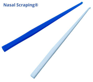
Nasal cytology testing involves the use of the single-use Nasal Scraping® device (which is significantly superior in terms of sample collection reliability and cell count) to collect the sample, May-Grunwald and Giemsa stains for MGG staining, and an optical microscope for the observation and digital acquisition of cytological images.

Nasal Cytology Sample Collection Using Nasal Scraping

Nasal swab sample collection for nasal cytology using Nasal Scraping – Rhinoscopy
VIDEO: Nasal cytology “fresh and fixed.”

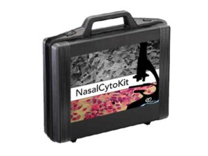
Nasalcytokit, nasal cytology
To learn more about nasal cytology, visit www.citologianasale.euorhttp://www.aicna.it.
Instructions for use will be available shortly.
Only customers who are logged in and have purchased this product can leave a review.
Related products
medical devices
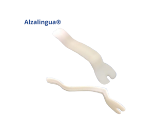
Alzalingua – Lingual Frenulum Device, 20-piece set
Price range: from €85.40 to €195.20 VAT included
Reviews
There are no reviews yet.